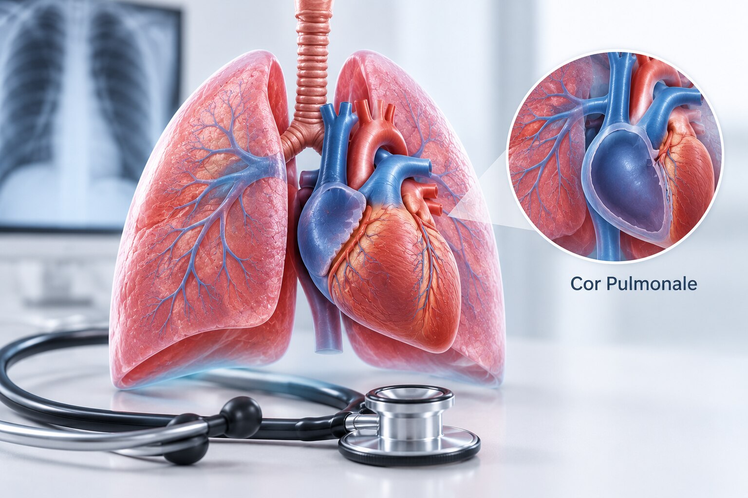
Breathlessness, falling oxygen levels and new swelling can signal that lung disease is placing sustained stress on the right side of the heart. You will learn which warning signs need urgent assessment, how a chest physician investigates the cause, what pulmonary function testing can and cannot show, and how to choose appropriate follow-up care.
Key takeaways
- Seek urgent care for severe breathlessness, chest pain, fainting, or blue lips.
- Ask what caused pulmonary hypertension before starting long-term treatment.
- Avoid smoking and report worsening swelling, fatigue, or reduced exercise tolerance.
- Follow test instructions and ask which pulmonary function results are reliable.
How lung disease creates right-heart strain
Cor pulmonale means enlargement or failure of the right ventricle after pulmonary hypertension develops from chronic lung disease, low oxygen or pulmonary-vessel disease. The right side must push blood through high-resistance lungs. This is different from left-sided heart failure, in which a weak left ventricle cannot pump effectively and can itself create pulmonary hypertension.
Ongoing right-heart strain may appear as reduced exercise tolerance, fatigue, ankle swelling, abdominal fullness, rapid fluid-related weight gain or needing extra pillows at night. These signs are not simply an isolated cough. A chest physician’s cor pulmonale assessment should investigate the source rather than label every breathlessness episode as heart failure.
| Condition | How it creates strain | What a chest physician may investigate |
|---|---|---|
| COPD and emphysema, including smoking heart lung disease | Persistent airflow limitation and low oxygen | Spirometry, full lung-function tests, oxygen assessment and chest imaging |
| Interstitial lung disease | Scarring reduces oxygen transfer | Diffusion capacity, high-resolution CT and exertional oxygen testing |
| Obstructive sleep apnoea | Repeated overnight oxygen drops | Sleep study and oxygen pattern |
| Recurrent pulmonary emboli | Blocked pulmonary vessels raise pressure | Clot history, CT pulmonary angiography or ventilation–perfusion scanning |
| Chest-wall restriction or neuromuscular weakness | Shallow breathing causes hypoventilation | Lung volumes, carbon-dioxide measurement and muscle-strength assessment |
Different causes need different treatments. Severe breathlessness at rest, fainting, chest pain, blue lips, confusion, coughing blood, or rapidly increasing swelling requires urgent assessment.
When lung strain becomes an urgent problem
Severe breathlessness at rest, fainting, chest pain, blue or grey lips, confusion, coughing blood, or a rapidly falling oxygen saturation needs emergency assessment, not a routine chest appointment. Rapidly increasing ankle or abdominal swelling is also urgent because it can signal decompensated right-heart failure.
- Breathlessness that suddenly becomes severe
- Oxygen saturation dropping quickly from your usual level
- New fainting, chest pain or coughing blood
- Blue or grey lips, unusual drowsiness or confusion
- Rapidly worsening ankle, leg or abdominal swelling
These changes can reflect an acute pulmonary embolism, infection, dangerous arrhythmia or worsening cor pulmonale. A chest physician assessing cor pulmonale needs to know how quickly the change occurred, because sudden deterioration carries a different risk from a gradual loss of walking ability.
| Pattern | What it suggests | What to do |
|---|---|---|
| Gradual reduction in walking distance over weeks | Progressive lung disease or increasing right-heart strain | Arrange prompt outpatient review |
| Sudden deterioration over minutes or hours | Pulmonary embolism, infection, arrhythmia or acute right-heart failure | Seek emergency assessment |
If you have an oximeter, record the number, pulse rate, time and symptoms, then bring the device if possible. Note when the change began and take your complete medication list. A single low reading needs context: warm your finger, sit still and check the signal quality.
Do not delay emergency care because the signal is poor when symptoms are severe.
What a chest physician checks before ordering treatment
A proper evaluation starts with the cause of breathlessness, not with a treatment label. The chest physician checks how far you can walk, when symptoms began, and whether they worsen during sleep or activity.
1. The history covers smoking, dust or chemical exposure at work, snoring, witnessed pauses in breathing, morning headaches, previous clots, chest infections, exacerbations, hospital admissions and every medicine, including inhalers, sedatives and non-prescription drugs.
2. Resting oxygen saturation is measured, then repeated during walking or another safe exertion. A poor signal or one isolated reading cannot establish chronic hypoxaemia.
3. Examination includes the chest and breathing pattern, heart sounds and rhythm, leg swelling, neck veins, liver enlargement and abdominal fluid. Existing spirometry, imaging, oxygen records and hospital reports should be reviewed rather than repeated automatically.
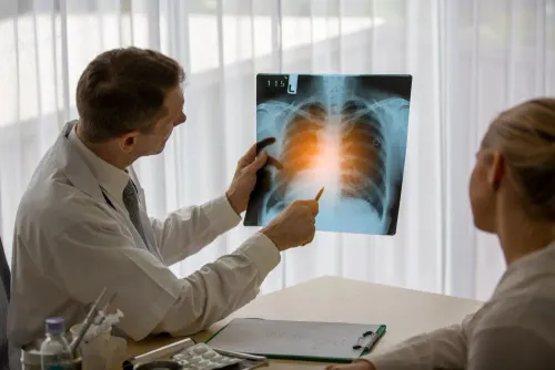
4. An ECG can show rhythm problems or right-heart strain. Chest X-ray or CT can reveal emphysema, interstitial disease, enlarged pulmonary arteries, infection or another explanation. A complete blood count checks anaemia and excess red cells; kidney and liver tests guide safe treatment, while BNP can support assessment of cardiac strain.
5. Echocardiography estimates pulmonary-hypertension probability and assesses right-ventricular size and function; it does not definitively measure pulmonary artery pressure. When confirmation will change management, right-heart catheterisation remains the haemodynamic reference standard.
The review should seek reversible stressors: untreated sleep-disordered breathing, ongoing smoking, anaemia, medication effects, undertreated COPD or asthma and recurrent thromboembolic disease. Severe, disproportionate or uncertain findings warrant cardiology or pulmonary-hypertension input. Search terms such as cor pulmonale lung care in Pune describe a service search, not a special diagnosis.
How to prepare for a pulmonary function test and read its limits
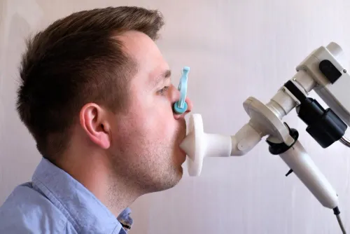
A pulmonary function test measures how air moves, how much your lungs hold, and how efficiently oxygen crosses into the blood; it does not diagnose cor pulmonale. Prepare for reliable results, then interpret them alongside heart and oxygen assessments.
- Avoid smoking beforehand and follow the clinic’s exact instructions about inhalers. Do not stop a prescribed inhaler unless the testing team tells you to.
- Wear loose clothing that will not restrict your chest or abdomen. Bring previous PFT reports, scans and medication details.
- Allow time for repeated forced-breathing manoeuvres. Spirometry records FEV1, FVC and the post-bronchodilator FEV1/FVC ratio; lung-volume testing detects restriction, while diffusion capacity (DLCO) assesses gas transfer.
- Read the result in context. A post-bronchodilator FEV1/FVC below 0.70 supports persistent airflow obstruction under the GOLD framework, but test quality, age, symptoms and the lower limit of normal still matter.
Restriction, obstruction or a low DLCO can reveal lung disease that strains the right heart, yet a ratio alone cannot diagnose or stage cor pulmonale. Resting and exertional oxygen levels, a timed walking test, imaging, ECG, echocardiography and blood tests complete the assessment; some patients need right-heart catheterisation.
Searches such as pulmonary function test cor pulmonale and chest physician cor pulmonale for pulmonary function test Hadapsar describe care needs, not special tests. The same applies to cor pulmonale lung care in Pune: confirm quality-controlled testing and coordinated cardiac evaluation.
Building a safe long-term plan for lung and right-heart strain
A safe outpatient plan targets the cause of lung and right-heart strain, not swelling alone. For smoking heart lung disease, compare clinics by whether they provide behavioural support and, when appropriate, nicotine replacement, varenicline or bupropion. Treat airway disease, sleep-disordered breathing and infection risks, and add pulmonary rehabilitation to improve strength and activity tolerance.
| Option | What to compare | When it applies |
|---|---|---|
| Oxygen assessment | Stable room-air PaO2 and saturation, measured reliably | Long-term oxygen is commonly considered at PaO2 ≤55 mm Hg or saturation ≤88%; breathlessness alone is not enough |
| Diuretic treatment | Weight, blood pressure, kidney function and electrolytes | Fluid-related ankle or abdominal swelling; monitoring prevents dehydration and electrolyte problems |
| Pulmonary-arterial-hypertension drugs | Confirmed cause and specialist assessment | Not automatic for COPD or interstitial lung disease |
| Respiratory testing | Quality-controlled spirometry with bronchodilator testing, full PFTs and oxygen assessment | Establishes treatment needs; PFT alone does not diagnose cor pulmonale |
A search for chest physician cor pulmonale for pulmonary function test Hadapsar should lead you to verify these capabilities, not assume that every clinic offers the same testing. For cor pulmonale lung care in Pune, also check whether the service coordinates echocardiography and pulmonary-hypertension referral when findings are severe or unclear.
Dr Kunjir's Brio Clinic can be considered alongside those checks when arranging respiratory follow-up in Pune. Keep scheduled reviews for oxygen readings, exercise tolerance, swelling, smoking status, inhaler technique, sleep treatment and rehabilitation progress.
Related service
Chest Diseases Treatment HEART DISEASES - COR PULMONALE Cor pulmonale is a condition that causes the right side of the heart to fail. View service → |
Frequently asked questions
How does chronic lung disease cause cor pulmonale?
Chronic lung disease, low oxygen, or pulmonary-vessel disease can raise resistance in the lung arteries. The right ventricle then works harder to move blood through the lungs, causing enlargement and eventual failure.
When is lung strain an urgent problem?
Seek urgent medical care for severe or rapidly worsening breathlessness, chest pain, fainting, confusion, blue lips, or sudden swelling. These symptoms can indicate dangerous heart or oxygen problems.
What does a chest physician check before treatment?
The assessment can include symptoms, smoking and medication history, oxygen saturation, chest examination, pulmonary function testing, blood tests, chest imaging, electrocardiography, and echocardiography. The tests depend on the suspected cause and severity.
How should you prepare for a pulmonary function test?
Follow the clinic's instructions about inhalers, smoking, heavy meals, alcohol, and strenuous exercise. Wear loose clothing, bring your medication list, and tell the technician if you feel dizzy or cannot maintain the required breathing effort.
Keywords