Chronic lung disease can reduce oxygen levels, increase pressure in the pulmonary circulation and force the right ventricle to work harder. A pulmonary function test helps identify the respiratory problem behind that strain, but it must be read alongside heart, oxygenation and imaging tests. By the end, you will know which PFT measurements matter, what their patterns suggest and when a broader chest-disease assessment is needed.
Key takeaways
- Spirometry identifies airflow obstruction but cannot confirm cor pulmonale alone.
- DLCO helps show impaired gas transfer linked to lung and pulmonary vascular disease.
- Combine PFT results with echocardiography, oxygen measurements and clinical findings.
- Avoid smoking and follow medication instructions before scheduled pulmonary testing.
How chronic lung disease can lead to right-heart strain
Long-standing lung disease can keep oxygen levels low, particularly during sleep or exertion. Low oxygen causes pulmonary arteries to constrict; persistent constriction raises pressure in the pulmonary circulation. The right ventricle then pumps against greater resistance, first thickening and later dilating or weakening. That progression is cor pulmonale.
A PFT helps identify the lung disorder behind this chain, but it does not measure pulmonary artery pressure or diagnose cor pulmonale. It can reveal airflow obstruction or restriction and impaired gas transfer. The lung function heart disease connection matters when clinicians decide whether the lung abnormality plausibly explains the right-heart strain.
DLCO adds an important clue: it measures how efficiently gas crosses from the air sacs into blood. A disproportionately low result can point toward pulmonary vascular disease, but it is not specific. Interpret it with haemoglobin, lung volumes and chest imaging; anaemia alone can lower DLCO.
In a pulmonary heart condition assessment, pair PFT findings with oxygenation and cardiac assessment. Echocardiography estimates the likelihood of pulmonary hypertension and examines right-ventricular size and function, while right-heart catheterisation confirms pulmonary pressures when definitive haemodynamic evidence is needed.
Severe PFT abnormalities do not automatically mean severe pulmonary hypertension, and modest spirometry does not exclude major vascular disease.
What each pulmonary function test adds
Each test answers a different question, so a pulmonary function test for cor pulmonale in Pune should usually include more than spirometry. These measurements identify the lung disorder contributing to pulmonary hypertension or right-heart strain; they do not measure pulmonary artery pressure or diagnose cor pulmonale alone.
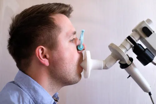
- Spirometry measures airflow, forced vital capacity and the forced expiratory volume in one second. A reduced FEV1/FVC ratio supports airway obstruction, as seen in COPD or asthma, while a low FVC raises concern for restriction but cannot confirm it without lung-volume testing.
- Bronchodilator testing repeats spirometry after an inhaled medicine. A meaningful improvement supports reversible airflow limitation, which points toward asthma or an asthma component; no improvement does not exclude symptoms, emphysema or pulmonary vascular disease.
- Lung volumes measure total lung capacity, residual volume and air trapping. A low total lung capacity confirms restriction from interstitial lung disease, chest-wall limitation or another cause, while increased residual volume can show trapped air that routine spirometry misses.
- DLCO measures how efficiently gas crosses from the air sacs into the blood. A disproportionately low result can point toward emphysema, interstitial lung disease or pulmonary vascular disease, but interpret it with haemoglobin, lung volumes and imaging.
A pulmonary function test cor pulmonale in Pune work-up therefore provides context rather than a final cardiac answer. Echocardiography assesses right-ventricular size and function, while right-heart catheterisation confirms pulmonary hypertension when definitive pressure measurements are needed.
How to read obstructive, restrictive and low-DLCO patterns
Start with the pattern, not a single number. Obstruction means narrowed airways: a reduced FEV1/FVC ratio supports COPD and can also occur with bronchiectasis. The ratio and FEV1 describe airflow limitation, but they do not measure pulmonary artery pressure.
1. Confirm restriction before interpreting it. A low FVC can result from air trapping, poor effort or true restriction. Reduced total lung capacity confirms restriction; a pattern with low lung volumes and reduced DLCO raises concern for interstitial lung disease, chest-wall limitation or another process outside the airways.
2. Check DLCO beside haemoglobin, lung volumes and imaging. A disproportionately low DLCO can point to emphysema, interstitial lung disease or pulmonary vascular disease, including pulmonary hypertension. It is a useful clue in pulmonary heart condition assessment, not a diagnosis.
3. Compare the abnormality with symptoms and oxygenation. Marked breathlessness, low oxygen or a very low DLCO with modest spirometric changes deserves attention because pulmonary vascular disease can be substantial even when airflow limitation is mild.
4. Treat bronchodilator response as context, not proof of cause. Improvement supports a reversible airway component, while a persistent abnormal pattern needs correlation with the clinical history.
PFTs explain the lung disease that may contribute to right-heart strain; they do not show right-ventricular enlargement or directly measure pulmonary pressure. Lung function heart disease assessment therefore requires oxygen testing, ECG, echocardiography and, when indicated, right-heart catheterisation. No single PFT value can prove cor pulmonale.
Why PFT results must be combined with heart and oxygen tests
PFT results must be combined with heart and oxygen tests because spirometry measures airflow, not pulmonary artery pressure, right-ventricular function or tissue oxygen delivery. A search for “pulmonary function test cor pulmonale in pune” should therefore lead to a broader assessment, not a diagnosis from one breathing result.
| Test | What it adds beyond spirometry | What an abnormal result may suggest |
|---|---|---|
| Echocardiogram | Estimates pulmonary pressure and shows right-ventricular size, wall thickness and pumping function | Pulmonary hypertension or right-heart strain |
| ECG | Records electrical patterns linked to right-ventricular overload | Right-axis deviation, right-heart strain or another cardiac cause |
| Pulse oximetry | Measures oxygen saturation at rest and during activity | Resting or exertional hypoxaemia that spirometry cannot detect |
| Arterial blood gas | Measures oxygen, carbon dioxide and blood acidity directly | Chronic hypoventilation, severe gas-exchange failure or respiratory acidosis |
| Six-minute walk test | Records walking distance, symptoms and oxygen desaturation | Functional limitation missed by resting tests; it is not specific for cor pulmonale |
| Right-heart catheterisation | Directly measures pressures in the right heart and pulmonary artery | Confirms pulmonary hypertension and defines its severity when non-invasive tests are inconclusive |
DLCO adds another clue by showing how efficiently oxygen crosses from the air sacs into blood. A very low value can point toward pulmonary vascular disease, emphysema or interstitial lung disease, but check it with haemoglobin, imaging and lung volumes.
Together, these results distinguish lung limitation from cardiac or oxygenation failure. That is the purpose of coordinated chest disease testing in pune: refine the cause, severity and urgency rather than overread one PFT.
Choosing and preparing for chest-disease testing in Pune
Check the test plan before you book. A useful cor pulmonale work-up may require spirometry with bronchodilator testing, lung volumes and DLCO, followed by oxygen assessment, ECG and echocardiography. Ask which tests are available, whether a respiratory physician will interpret them together, and whether you should bring previous scans, prescriptions and discharge records.
At Dr Kunjir's Brio Clinic, this question helps prevent treating a PFT report as a diagnosis on its own.
- Tell the testing centre about an active cough with fever or a contagious respiratory infection; postponing protects staff and prevents unreliable effort during illness.
- Report eye surgery, chest surgery, unstable heart disease, recent hospitalisation or an inability to perform forceful breathing manoeuvres.
- Ask how to take inhalers before testing. Do not stop a prescribed medicine unless the clinician gives you a specific instruction.
- Wear loose clothing, avoid smoking before the appointment, and ask whether a six-minute walk test will be added to record exertional oxygen desaturation.
| Situation | What to do | Why |
|---|---|---|
| Stable symptoms and safe forced breathing | Arrange elective chest-disease testing in Pune | PFT is usually a low-risk outpatient assessment |
| Active contagious infection, recent eye or chest surgery, or unstable heart disease | Call the centre before attending | You may need postponement or modified testing |
| Fainting, chest pain, severe resting breathlessness or suspected acute cor pulmonale | Seek urgent medical assessment, not an elective slot | Waiting for PFT could delay treatment of a dangerous heart-lung problem |
Frequently asked questions
How can chronic lung disease cause cor pulmonale?
Persistent low oxygen can constrict pulmonary arteries, raise pulmonary circulation pressure and force the right ventricle to pump harder. Over time, the ventricle can thicken, dilate or weaken.
What does each pulmonary function test add to cor pulmonale assessment?
Spirometry measures airflow, lung-volume testing identifies restriction or hyperinflation, and DLCO measures gas transfer across the lungs. Together, they help define the lung problem contributing to right-heart strain.
How are obstructive, restrictive and low-DLCO patterns interpreted?
An obstructive pattern shows reduced airflow, a restrictive pattern shows reduced lung volumes, and a low DLCO shows impaired gas transfer. Clinicians interpret these patterns with symptoms, imaging and heart tests.
Why are PFT results combined with heart and oxygen tests?
PFTs describe lung mechanics and gas transfer but do not measure pulmonary artery pressure or right-heart function. Echocardiography, oxygen saturation, blood-gas testing and exercise assessment provide complementary evidence.
How should you prepare for chest-disease testing in Pune?
Bring previous reports and your medication list, follow instructions about inhalers, avoid smoking before testing and wear comfortable clothing. Ask the testing centre whether you need to withhold a specific inhaler.
Keywords