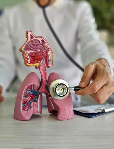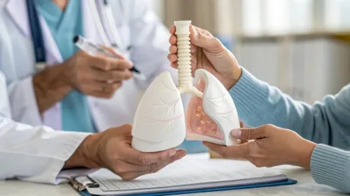
A pulmonary function test measures how well air moves through and out of your lungs, but it cannot confirm bronchiectasis by itself. You will know which tests answer which clinical questions, how to prepare, and what to ask when symptoms continue despite a normal result.
Key takeaways
- Spirometry measures airflow but cannot confirm permanent bronchial widening.
- Prepare for spirometry by avoiding smoking and following medicine instructions from your clinic.
- HRCT confirms airway changes; sputum culture identifies bacteria that treatment may need to target.
- Compare testing by specialist review, same-day access, written results and follow-up planning.
Which tests are used to assess suspected bronchiectasis?
Suspected bronchiectasis is assessed by combining daily cough, sputum, recurrent infections, examination findings, prior pneumonia or tuberculosis and objective testing. Spirometry is usually the starting respiratory test. It records forced expiratory volume in one second (FEV1), forced vital capacity (FVC) and the FEV1/FVC ratio, showing airflow impairment rather than permanent airway widening.
A pulmonary function test for bronchiectasis cannot diagnose the condition alone. Confirmation requires compatible clinical findings plus permanent bronchial dilatation on thin-section or high-resolution CT (HRCT) of the chest. A normal chest X-ray or near-normal spirometry does not reliably exclude it.
The wider assessment may include:
- Bronchodilator reversibility testing, which checks whether airflow improves after an inhaled medicine.
- Lung volumes, including total lung capacity and residual volume, to identify restriction or air trapping.
- Diffusion capacity for carbon monoxide (DLCO), which assesses gas transfer.
- Pulse oximetry at rest and a six-minute walk test for exertional oxygen change, distance and symptoms.
- Sputum culture, preferably a lower-airway sample collected before antibiotics when possible.
| Test | Main role | What it cannot establish |
|---|---|---|
| Spirometry | Measures airflow | Permanent airway widening |
| HRCT chest | Shows bronchial dilatation and structural changes | Day-to-day airflow performance |
| Six-minute walk test | Measures functional exercise response | A substitute for CT or spirometry |
Bronchiectasis respiratory testing in Pune should therefore be interpreted as a set of results, not one decisive number.
What happens during spirometry, and how should you prepare?
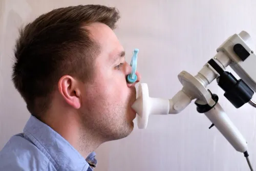
You usually sit upright for spirometry, wearing a nose clip and sealing your lips around a disposable mouthpiece. In this pulmonary function test for bronchiectasis, you inhale fully, then blow out as hard and as long as instructed.
1. The technician repeats the manoeuvre several times to check whether FEV1, FVC and the FEV1/FVC ratio are acceptable and repeatable. Coughing, stopping early, failing to inhale fully, leaking around the mouthpiece or giving inconsistent effort can produce a falsely low or uninterpretable result.
2. A bronchodilator may be given before repeat blows. This helps identify reversible airflow limitation or coexisting asthma, but a response does not prove bronchiectasis, and no response does not rule it out.
3. Ask the testing centre for specific instructions before stopping any prescribed inhaler or other medicine. Also ask when to avoid smoking, vigorous exercise, a large meal and caffeine, because medication effects and test timing can alter results.
4. Allow about 15 to 30 minutes for spirometry. It is not painful, but repeated maximum-effort blows can leave you briefly light-headed or tired; tell the technician if that happens.
Complete PFTs take longer when lung volumes or DLCO are included. This preparation matters whether you arrange respiratory testing in Pune or elsewhere, because reliable technique is essential when assessing symptoms linked to bronchiectasis.
How are PFT, oxygen and exercise results interpreted?
Interpretation depends on the pattern, test quality and change over time, not one number. Obstruction appears as a reduced FEV1/FVC ratio. Air trapping may require lung-volume measurement: a raised residual volume suggests trapped air, while a reduced total lung capacity confirms restriction when spirometry suggests it. Mixed obstruction and restriction can occur.
| Test | What it measures | What the result means |
|---|---|---|
| Spirometry | FEV1, FVC and FEV1/FVC | Reduced ratio supports obstruction; preserved airflow does not exclude structural bronchiectasis |
| Lung volumes | Residual volume and total lung capacity | Detects air trapping and confirms restriction when total lung capacity is reduced |
| DLCO | Gas transfer from air sacs to blood | Investigates breathlessness exceeding spirometry findings or another coexisting lung disorder |
| Pulse oximetry | Oxygen saturation at rest | Shows resting oxygenation, not exercise limitation |
| Six-minute walk test | Distance, symptoms, heart rate and oxygen saturation during exertion | Measures function; it does not replace HRCT or spirometry |
A report should state whether blows were acceptable and repeatable. Compare stable-state tests made with similar equipment and quality standards. A fall during an acute flare can reflect infection, mucus plugging or poor effort, so one abnormal result does not prove permanent progression.
Smoking can worsen mucus and lung-function decline, but smoking does not establish a bronchiectasis diagnosis. Persistent symptoms or repeated infections need clinical review even when airflow measurements remain preserved, because structural airway damage and physiological impairment do not always match.
Why HRCT and sputum culture often matter more than a single PFT
Thin-section HRCT is the key test for confirming bronchiectasis because it shows permanent airway widening that spirometry cannot see. Radiologists look for a bronchus wider than its accompanying artery, absent normal bronchial tapering and bronchi visible close to the pleura.
HRCT identifies structure; a PFT measures airflow, lung volumes and gas transfer. A single PFT can look near-normal despite bronchiectasis, so it cannot establish or exclude the diagnosis on its own.
Stable-state sputum should be collected before antibiotics when possible. A lower-airway sputum sample is more useful than a throat swab when you can produce sputum. Repeated cultures guide future antibiotic choices and identify organisms such as Pseudomonas aeruginosa; producing no sputum does not prove that infection or bronchiectasis is absent.
A practical work-up also searches for treatable causes and associations:
- Request a full blood count and immunoglobulins IgG, IgA and IgM.
- Check total IgE and Aspergillus sensitisation for allergic bronchopulmonary aspergillosis.
- Add mycobacterial testing when clinical features indicate it.
- Record smoking exposure, because smoking can worsen mucus, cough and lung-function decline or coexist with COPD.
A smoking bronchiectasis diagnosis still requires objective assessment. Persistent daily sputum, recurrent infections or unexplained breathlessness should not be attributed automatically to tobacco exposure.
How to choose a bronchiectasis assessment in Pune
Choose a respiratory service that treats the assessment as a whole, not as a spirometry appointment alone. Ask whether the clinician will review the actual HRCT images, not only the report; compare prior spirometry; and check that the PFT report states acceptability and repeatability.
| Assessment | Ask for | When it helps |
|---|---|---|
| Spirometry | FEV1, FVC, FEV1/FVC and quality comments | Baseline airflow comparison |
| Complete PFT | Lung volumes and DLCO | Breathlessness unexplained by spirometry |
| Six-minute walk test | Distance, symptoms, heart rate and oxygen saturation | Exertional limitation |
| Sputum culture | Lower-airway sample and antibiotic timing | Daily sputum or recurrent infections |
| HRCT review | Direct image review by the clinician | Confirming structural disease and extent |
Bring these records and details:
- Prior pneumonia or tuberculosis records, recurrent infection dates, asthma or COPD diagnoses, smoking exposure, and medicines.
- Aspiration or swallowing concerns, autoimmune disease, sputum culture reports, and vaccination history.
- Ask when to test during a stable period and whether sputum should be collected before antibiotics.
A fall during an acute flare can reflect infection, mucus plugging or poor effort, so track change with comparable stable-state tests. For someone searching for a bronchiectasis chest physician in Pune, Dr Kunjir's Brio Clinic can be one option for a consultation linking symptoms, CT findings, PFT quality, sputum results and possible causes.
Seek prompt medical assessment for coughing blood, severe breathlessness, chest pain, confusion or blue lips. The useful outcome is a plan to confirm the diagnosis, identify infection or a treatable association, and monitor change—not an isolated spirometry number.
Related service
Chest Diseases Treatment Asthma triggered by exposure to the same substances that trigger allergy symptoms. View service → |
Frequently asked questions
Which tests assess suspected bronchiectasis?
Assessment can include spirometry or full PFT, pulse oximetry, exercise oxygen testing, HRCT chest and sputum culture. Your symptoms, examination and history determine the combination.
How should you prepare for spirometry?
Follow the clinic’s instructions about inhalers, avoid smoking before the test, wear comfortable clothing and tell the technician about recent illness or chest pain.
What do PFT, oxygen and exercise results show?
PFT results show airflow and lung volumes, while oxygen and exercise tests show whether oxygen levels fall or breathlessness increases with activity. Results need clinical interpretation.
Why can HRCT and sputum culture matter more than one PFT?
HRCT can show widened or damaged airways, which spirometry cannot confirm. Sputum culture can identify bacteria and guide antibiotic selection.
How do you choose bronchiectasis respiratory testing in Pune?
Choose a centre that can arrange the required tests, explain preparation, provide clear reports and offer review by a chest physician who can connect the findings to your symptoms and history.
Keywords